Semisynthesis of Stable Isotope-Labeled Ergot Alkaloids for HPLC-MS/MS Analysis

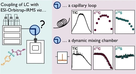
J. Agric. Food Chem. 2025, 73, 29, 18412–18419: Graphical abstract
Ergot alkaloids (EAs) are common contaminants in cereals such as rye, wheat, and barley, posing food safety challenges worldwide. Their quantification, according to EN 17425:2021, relies on HPLC-MS/MS analysis of six primary EAs and their epimers. However, accurate quantification is often hindered by complex food matrices and the lack of suitable internal standards.
To overcome these limitations, a semisynthetic approach was developed to produce stable isotope-labeled analogs of all major EAs. Building on previous work, native ergotamine was N6-demethylated and remethylated using ^13CD₃-iodomethane. The resulting labeled standards were validated against native compounds using HPLC-HR-MS/MS, exhibiting identical chromatographic and mass spectral properties. These new standards significantly improve accuracy, reproducibility, and comparability of EA analyses, strengthening food safety monitoring.
The original article
Semisynthesis of Stable Isotope-Labeled Ergot Alkaloids for HPLC-MS/MS Analysis
Sven-Oliver Herter, Hajo Haase, and Matthias Koch*
J. Agric. Food Chem. 2025, 73, 29, 18412–18419
https://doi.org/10.1021/acs.jafc.5c03345
licensed under CC-BY 4.0
Selected sections from the article follow. Formats and hyperlinks were adapted from the original.
Ergot alkaloids (EAs) are a group of naturally occurring toxic alkaloids produced by fungi of the genus Claviceps, with Claviceps purpurea being the most prevalent in Europe. (1) They grow on various cereals, but primarily on rye, wheat, and triticale. (2,3) When a floret is infected with a spore of ergot fungi, the spore mimics a pollen grain growing into the ovary during fertilization, thus destroying the plant ovary and connecting to the vascular bundle of the plant. The ergot fungi produce asexual spores that are released with a sugary honeydew and dispersed by insects to other florets. In preparation for winter, a hardened mycelium (sclerotia) is formed inside the husk of the floret, and alkaloids and lipids are accumulated in the sclerotium. (4) The harvest of infected cereal leads to the introduction of sclerotia into the food chain. Mechanical techniques for the removal of sclerotia from grain are effective; however, they do not eliminate contamination completely. Residual sclerotia, in the form of dust or fragments, are ground with grain to flour and thereby introduce toxic EAs into foodstuffs. (4,5) A defining characteristic of EAs is the tetracyclic ring structure, known as ergoline (Figure 1). Naturally occurring EAs diverge structurally from ergoline due to the methylation of the N6-atom and various substituents attached to the C8-atom. (6) Additionally, most EAs possess a double bond between the C9- and C10-atoms in the ergoline structure. (7) They can be divided into three different groups based on their structural characteristics: lysergic acid and its simple amides, the ergopeptines, and the ergoclavines. (6) They all share an R-configuration at the C8-position of the ergoline structure. Epimerization at the C8-atom of simple lysergic acid amides and ergopeptines via keto–enol tautomerism is observed when these EAs are exposed to heat, UV light, or high/low pH. (8,9) However, ergoclavines, such as lysergine and lysergole, do not undergo epimerization due to the absence of a carboxyl group at the C8-atom.
In parallel with the establishment of the maximum levels, the European Standard Method (ESM) EN 17425 for the determination of ergot alkaloids in cereals and cereal-based products was published. (14) The method describes the extraction of EAs from cereals and the subsequent cleanup of the extract using dispersive solid-phase extraction. The determination of EAs is performed with high-performance liquid chromatography coupled to tandem mass spectrometry (HPLC-MS/MS). However, the European Committee for Standardization (CEN) has indicated that the method cannot be considered satisfactory for levels below 24 μg/kg for the sum of the priority EAs due to an increase in interlaboratory uncertainties observed during the method validation study. (14) HPLC-MS/MS is susceptible to various forms of interference when used for quantification. A common challenge is the effect of coeluting molecules on the ionization yield of the analyte molecules. These matrix-dependent signal suppression or enhancements impact the analyte signal and cause the measured concentration to be different from its true value. (15,16) To overcome this problem, matrix-matched calibration can be employed. This involves the use of a calibration solution whose matrix matches the sample matrix, except for the analyte. However, these specific matrices are rarely available in environmental and food analytical studies. Alternatively, an internal standard (ISTD) can be used to reduce the impact of the matrix on the analyte signal. A suitable ISTD has comparable physio-chemical properties to the analyte, e.g., retention time, ionization response, and fragmentation pattern. Because of the need for an appropriate ISTD for each analyte of interest, this approach is costly and time-consuming. Therefore, in HPLC-MS/MS, ISTDs are typically stable isotope-labeled analogs (2H, 13C, and 15N) of the analyte. (17) For EAs, only ergometrine-13CD3 and its epimer ergometrinine-13CD3 are currently commercially available, due to their less complex structure compared to the ergopeptides. (15,18) However, a comprehensive set of all isotopically labeled priority EAs is required to enhance the current ESM and thus to reliably control the EA maximum level in food for infants and young children at 20 μg/kg. In our previous work, we described a straightforward two-step semisynthesis of the ergopeptines 13CD3-ergotamine and 13CD3-ergotaminine, starting from native ergotamine. (19) The general approach for the two-step semisynthesis of isotopically labeled EAs is illustrated in Figure 2. In the first step, native EA is demethylated at the N6-position via an iron-catalyzed dealkylation reaction to yield the corresponding Norergot alkaloid (Nor EA). Subsequent methylation at the N6-atom with an isotopically labeled methylating reagent, e.g., iodomethane, yields the isotopically labeled EA.
Given that all priority EAs share the methyl group at the N6-atom, we investigated the possibility of synthesizing all isotopically labeled EAs via a two-step synthesis.
Materials and Methods
HR-MS/MS Measurements
High-resolution tandem mass spectra (HR-MS/MS) were measured on a Quadrupole time-of-flight 6600 mass spectrometer (Sciex, Darmstadt, Germany). The purified solution, comprising either the native EA or Nor EA, was injected directly into the mass spectrometer via a syringe pump for analysis. For HPLC-HR-MS/MS analysis, the QTOF was coupled to a 1290 Infinity II system (Agilent, Waldbronn, Germany). For the measurements, a Phenomenex Gemini C6–Phenyl (150 × 2.0 mm; 3 μm) column was used. For the MS/MS experiments, an inclusion list with the exact m/z ratio [M + H]+ for the native and isotopically labeled EAs was created. Table S6 presents the QTOF parameters, and Table S7 presents the HPLC parameters that were used for the HPLC-HR-MS/MS measurement. The predicted sum formulas for the most abundant product ion m/z ratios were calculated using a threshold of ±5 ppm deviation (Tables S9–S14).
Results
HR-MS/MS of Norergot Alkaloids
The successful N6-demethylation of the EAs to the Nor EAs was confirmed by HR-MS/MS. Purified samples containing either the native EA or the associated Nor EA were introduced into the QTOF mass spectrometer for measurement via direct infusion. The HR-MS/MS spectra of the native EAs and Nor EAs were compared, and specific fragment ions were assigned to the major peaks in the mass spectra. Figure 4 shows the comparison of the fragment spectra of native ergocristine and the demethylated product norergocristine. The HR-MS/MS spectra of ergocristine coincide with previously reported data in the literature. (20,21) Upon comparison of the spectra of ergocristine and norergocristine, a similar fragmentation pattern is evident. The observed differences in the intensities of certain fragment ions can be attributed to the lower energy required for the fragmentation of norergocristine. For fragment ions containing the N6-atom, a shift in the m/z ratio for norergocristine compared to ergocristine was observed. This m/z shift corresponds to the loss of the methyl group at the N6-atom and is observed for the fragment ions of ergocristine/norergocristine with the m/z ratios of 348.1711/334.1545, 268.1451/254.1280, and 223.1234/209.1073. The differences in the m/z are 14.0166, 14.0171, and 14.0164, respectively, and match with the m/z ratio of the neutral loss of CH2, which is 14.0157 (cf. Figure 4).
![J. Agric. Food Chem. 2025, 73, 29, 18412–18419: Figure 4. Positive ESI-HR-MS/MS spectra [M + H]+ of (a) ergocristine and (b) norergocristine. In addition to the measured accurate m/z ratio, a suggestive structure is provided for the precursor ion and the major fragment ions along with their calculated exact m/z ratio.](https://lcms.labrulez.com/labrulez-bucket-strapi-h3hsga3/J_Agric_Food_Chem_2025_73_29_18412_18419_Figure_4_Positive_ESI_HR_MS_MS_spectra_M_H_of_a_ergocristine_and_b_norergocristine_3403f0b9e7_l.webp) J. Agric. Food Chem. 2025, 73, 29, 18412–18419: Figure 4. Positive ESI-HR-MS/MS spectra [M + H]+ of (a) ergocristine and (b) norergocristine. In addition to the measured accurate m/z ratio, a suggestive structure is provided for the precursor ion and the major fragment ions along with their calculated exact m/z ratio.
J. Agric. Food Chem. 2025, 73, 29, 18412–18419: Figure 4. Positive ESI-HR-MS/MS spectra [M + H]+ of (a) ergocristine and (b) norergocristine. In addition to the measured accurate m/z ratio, a suggestive structure is provided for the precursor ion and the major fragment ions along with their calculated exact m/z ratio.
The fragmentation of the amide bond between the lysergic acid moiety and the cyclic tripeptide corresponds to the fragment ions with m/z ratios of 223.1234/209.1070 and 268.1451/254.1280. Fragmentation of the cyclic tripeptide ring results in the fragment ions with m/z ratios of 348.1711/334.1545. The fragment ion with an m/z ratio of 325.1546 is indicative of the fragment ion of the tricyclic peptide. Given that no modifications were introduced to the cyclic tripeptide ring structure, the spectra of ergocristine and norergocristine both exhibit this specific fragment ion. The HR-MS/MS spectra for the Nor EAs of ergometrine, ergocornine, ergotamine, α-ergocryptine, and ergosine compared to the native EAs are presented in the Supporting Information, Figures S7, S9, S11, S13, S15. The related structure of all investigated ergopeptines results in a comparable fragmentation pattern, similar to the one observed for ergocristine and norergocristine. In contrast, the representatives of the lysergic acid derivatives, ergometrine and norergometrine, exhibit a less complex product ion spectrum. Based on the measured accurate m/z ratios, we were able to make a structural proposal for all major product ions of the EAs and Nor EAs.
Synthesis of Stable Isotope-Labeled Ergot Alkaloids
To verify the successful synthesis of all 12 isotopically labeled priority EAs, they were spiked at a concentration of 25 ng/mL to a standard with a concentration of 25 ng/mL containing all unlabeled native EAs in acetonitrile and analyzed via HPLC-HR-MS/MS. Figure 5 depicts the extracted-ion chromatogram (XIC) for the specific fragment ion of the native (m/z 223.1235) and isotopically labeled (m/z 227.1458) EAs. (22)
![J. Agric. Food Chem. 2025, 73, 29, 18412–18419: Figure 5. Extracted-ion chromatogram [M + H]+ (XIC) of a 25 ng/mL standard for (a) 12 native priority EAs and (b) 12 stable isotope-labeled priority EAs. An inclusion list containing the exact m/z values of each native and stable isotope-labeled EA [M + H]+ was created, and the sample was analyzed in parallel reaction monitoring (PRM) mode on a QTOF mass spectrometer.](https://lcms.labrulez.com/labrulez-bucket-strapi-h3hsga3/J_Agric_Food_Chem_2025_73_29_18412_18419_Figure_5_Extracted_ion_chromatogram_M_H_XIC_of_a_25_ngm_L_standard_70f2a2760a_l.webp) J. Agric. Food Chem. 2025, 73, 29, 18412–18419: Figure 5. Extracted-ion chromatogram [M + H]+ (XIC) of a 25 ng/mL standard for (a) 12 native priority EAs and (b) 12 stable isotope-labeled priority EAs. An inclusion list containing the exact m/z values of each native and stable isotope-labeled EA [M + H]+ was created, and the sample was analyzed in parallel reaction monitoring (PRM) mode on a QTOF mass spectrometer.
J. Agric. Food Chem. 2025, 73, 29, 18412–18419: Figure 5. Extracted-ion chromatogram [M + H]+ (XIC) of a 25 ng/mL standard for (a) 12 native priority EAs and (b) 12 stable isotope-labeled priority EAs. An inclusion list containing the exact m/z values of each native and stable isotope-labeled EA [M + H]+ was created, and the sample was analyzed in parallel reaction monitoring (PRM) mode on a QTOF mass spectrometer.
Discussion
The quantification of EAs in complex food matrices using HPLC-MS/MS is challenging, primarily due to matrix effects and the lack of stable isotope-labeled standards. To address this gap, a semisynthetic approach was developed that targets the N6-atom of the lysergic acid moiety, a structural feature shared among all investigated native priority EAs.
The demethylation of the N6-atom to the corresponding Nor EAs was achieved via an iron-catalyzed reaction, and the structure of the Nor EAs was confirmed by HR-MS/MS. A structural assignment for the most intense product ions of the Nor EAs was proposed based on the measured accurate m/z ratios and correlated with specific HR-MS/MS fragments of the native compounds. The characteristic loss in the m/z value associated with the demethylation was observed only for fragment ions of the Nor EAs that contain the N6-atom, thereby confirming successful and selective demethylation for all investigated EAs.
For the subsequent remethylation of the Nor EAs, 13CD3-iodomethane was utilized and resulted in the formation of the isotopically labeled C8-R and C8-S epimers of the respective EAs. Overall yields ranged from 8.1% for 13CD3-ergocristine/-inine and up to 29.5% for 13CD3-ergocryptine/-inine. Due to the limited availability of starting material, no yield could be determined for 13CD3-ergometrine/-inine and 13CD3-ergosine/-inine. The epimer ratio (R%:S%) of the ergopeptines ranged between 58:42 for 13CD3-ergocornine/-inine and 45:55 for 13CD3-ergocristine/-inine. Although the synthesis started from a compound with the 8R-configuration, both stable isotope-labeled epimers were obtained in approximately equal amounts. This outcome is favorable as it provides sufficient quantities of both stable isotope-labeled epimers from a single epimeric pure starting material. In contrast, the simpler lysergic acid amide 13CD3-ergometrine/-inine showed less epimerization, with an epimer ratio of 69:31. The structures of the synthesized labeled EAs were confirmed using HPLC-HR-MS/MS. Therefore, a mixture containing all 12 native priority EAs and their corresponding labeled analogs was analyzed. As anticipated for deuterium-labeled compounds, the isotopically labeled EAs showed slightly shorter retention times (1–3 s) compared to their native counterparts. The precursor and fragment ions containing the isotopically labeled methyl group at the N6-position exhibited a shift in the m/z ratio of 4 Da. In contrast, fragment ions without this structural feature were present in the MS/MS spectra of both labeled and unlabeled EAs. With the measured accurate m/z ratios of the precursor and product ions, we were able to predict a sum formula and match specific fragment ions for each major signal in the HR-MS/MS spectrum. Due to the limited availability of the native EAs, only small amounts of the 13CD3-labeled analogs were synthesized, which precluded structural elucidation by NMR. However, the identical fragmentation patterns and signal intensities between labeled and unlabeled EAs, in conjunction with the same retention time in HPLC, provided unambiguous confirmation of the structures for all 12 synthesized stable isotope-labeled priority EAs.
For the first time, the procedure described herein enabled the semisynthesis of a complete set of stable isotope-labeled priority EAs. In future work, these standards will be implemented in the ESM and facilitate an enhanced methodology for the quantification of these mycotoxins in food and feed.