Residue-Level Determination of Small-Molecule–Protein Affinities by Hydrogen–Deuterium Exchange Mass Spectrometry
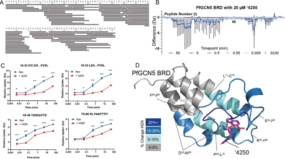
J. Am. Soc. Mass Spectrom. 2026, 37, 6, 1391–1401: Figure 1. Global shifts in solvent exchange in PfGCN5-BRD on compound binding. (A) Peptide map of PfGCN5-BRD showing coverage of pepsin-produced peptide map. (B) Global HDX profile of PfGCN5-BRD on incubation with 20 μM ′4250. Each time point is shown alongside the sum of the shifts (SUM). (C) Representative peptide uptake plots of four peptides showing changes in ′4250 binding. Each data point is the average with standard deviation shown as error bars (n = 3). * = > 5% and >0.5 Da difference and passes a student t-test with p < 0.05, **p < 0.01, and ***p < 0.001. (D) Shifts in HDX mapped onto the PfGCN5-BRD:′4250 cocrystal structure, solved to 1.8 Å resolution. PDB: 9TM1.
This study presents an advanced hydrogen–deuterium exchange mass spectrometry (HDX-MS) workflow that enables quantitative determination of small-molecule–protein binding affinities with residue-level resolution. By combining HDX-MS/MS, electron capture dissociation, and ligand titration experiments, the method estimates apparent dissociation constants at the global, peptide, and individual amino acid levels.
The approach provides spatially resolved affinity measurements while taking advantage of automated HDX-MS workflows for sample handling and data analysis. It offers a scalable platform for mapping ligand–protein interactions and supports structure–activity relationship studies in drug discovery and biopharmaceutical research.
The original article
Residue-Level Determination of Small-Molecule–Protein Affinities by Hydrogen–Deuterium Exchange Mass Spectrometry
De Lin, Luma Godoy Magalhaes, Joel McMillan, Thomas C. Eadsforth, Greg Stewart, Kieran R. Cartmill, Vincent L. G. Postis*, and Glenn R. Masson*
J. Am. Soc. Mass Spectrom. 2026, 37, 6, 1391–1401
https://doi.org/10.1021/jasms.6c00020
licensed under CC-BY 4.0
Selected sections from the article follow. Formats and hyperlinks were adapted from the original.
Hydrogen–Deuterium Exchange Mass Spectrometry (HDX-MS) has emerged as a key technique in structure-based drug discovery, enabling a rapid characterization of protein–ligand interactions and conformational dynamics in both biopharmaceutical and small molecule development. (1−8) Typically, HDX-MS studies follow a “bottom-up” workflow in which the therapeutical target is incubated with a saturating concentration of ligand in the presence of a deuterated buffer for a series of defined time points. Hydrogen–deuterium exchange is then quenched, and the protein chemically denatured. The denatured protein is then proteolytically digested, and the resulting peptides are analyzed by LC–MS to determine the extent of deuterium incorporation across their sequences. (9) These resulting data provide peptide-level spatial resolution, typically 5–35 amino acids, allowing the identification of regions stabilized or shielded from the solvent upon ligand binding. (10) Higher resolution, including single-residue resolution, can be achieved computationally, especially when high peptide redundancy and sequence coverage is obtained. (11−13)
To minimize heterogeneous exchange behavior, ligand-binding HDX-MS experiments are commonly conducted at concentrations well above the compound dissociation constant (KD), producing a fully bound complex to avoid heterogeneous bimodal isotopic envelopes. (2) This belies the fact that HDX-MS workflows have been previously described which are capable of determining affinity constants, such as PLIMSTEX (14) and SUPREX, (15) and a push to make HDX-MS more “quantitative”, i.e., able to abstract meaningful (and crucially comparable) biophysical constants from HDX-MS data. (16) Furthermore, there has been renewed interest, notably from the Wilson group and co-workers, in HDX-MS/MS (i.e., the fragmentation of a deuterated precursor peptide to produce deuterated product ions), which enables residue-level investigation of deuterated peptides. (17−19) These experiments build on the comprehensive and groundbreaking work in the groups of Jørgensen and Rand, who used electron capture dissociation (ECD) or electron transfer dissociation (ETD) to achieve fragmentation with minimal hydrogen/deuterium (H/D) scrambling. (20−23) Although HDX-MS/MS has been applied to ligand binding, (24,25) its use has remained limited, possibly due to the low efficiency of fragmentation and the stringent controls required to detect signal scrambling. Newer technologies, such as Electron-Activated Dissociation (EAD), have shown improved performance, increasing the accessibility of scramble-free HDX-MS/MS for ligand screening. (17)
Here, we report a compound titration experiment using both HDX-MS and ECD-based HDX-MS/MS that enables the determination of apparent dissociation constants (KDapp) with an amino acid spatial resolution. Using the Plasmodium falciparum GCN5 bromodomain as a model system, we measured deuterium uptake as a function of compound concentration at the peptide and single-amino acid level to extract quantitative affinity information. Comparison with orthogonal biophysical methods showed a strong agreement between HDX-derived and reference KD values. This approach provides a time-efficient, low-sample-consumption, in-solution method of quantifying ligand binding and mapping local conformational changes, enabling detailed characterization of the mode of interaction and binding mechanisms in protein–ligand events.
Methods
Hydrogen–Deuterium Exchange-Mass Spectrometry
HDX-MS data were acquired using the HDX manager (Waters Corp.) maintained at 0.1 °C and coupled in-line to a SELECT SERIES Cyclic IMS QTOF mass spectrometer (Waters Corp.). Automated liquid handling was performed using a PAL3 robotic tool change system, controlled by Chronos Software (LEAP Technologies). The protein was digested in-line for 2 min at 20 °C using an Enzymate BEH Pepsin 2.1 mm × 30 mm column (Waters Corp.). The generated peptides were desalted in buffer A (0.1% formic acid in water) using an ACQUITY UPLC BEH C18 VanGuard Precolumn (1.7 μm, 5 × 2.1 mm; Waters Corp.) prior to reversed-phase separation on an ACQUITY UPLC C18 column (1.7 μm, 100 × 1 mm; Waters Corp.). Peptides were eluted using a 40 μL/min flow rate from 5% to 85% buffer B (0.1% formic acid in acetonitrile) in buffer A. Data were acquired in positive mode over a m/z range of 50–1200 with a spray voltage of 2.0 kV with leucine enkephalin (m/z 556.2766) used as a lockmass internal calibrant during data acquisition.
Results and Discussion
Dissociation Constants Determined via Peptide-Level HDX-MS/MS Analysis
We next sought to determine whether ECD could be used under minimized H/D-scrambling conditions to determine apparent dissociation constants (KDapp) at single-amino-acid resolution. The source and ECD cell parameters were optimized for minimal deuterium scrambling, initially using the P1 peptide HDX scrambling monitor to tune as previously described (21,24,37,42) with further refinement being conducted using the loss of ammonia method as described by Rand et al. (22) (Supporting Information Figure S1). Targeted ECD fragmentation was then applied to deuterated precursor ions of the PfGCN5-BRD peptide containing residues 18 to 33. This peptide, detected predominantly in +3 charge state and with a relative abundance exceeding 105 counts, produced an almost complete c ion series including c3-c7 and c9-c11 and z ion fragments z9-z13, providing a near-complete single-residue coverage (Figure 3).
![J. Am. Soc. Mass Spectrom. 2026, 37, 6, 1391–1401: Figure 3. Representative ECD MS/MS spectra of the [M + 3H]3+ precursor ion of the peptide LEKQQSAWPFLKPVSL acquired under nondeuterated and deuterated conditions. (A) ECD spectrum of the nondeuterated peptide at 0 s exchange (precursor m/z 624.40, 3+) and ECD spectrum following 5 min deuterium labeling (precursor m/z 626.30, 3+). Spectra are displayed at the indicated relative intensity scales. Predominantly c- and z-type ions consistent with nonergodic ECD cleavage were observed. (B) High-resolution isotopic distributions of a representative ECD fragment ion (c11) of the peptide LEKQQSAWPFLKPVSL under nondeuterated and deuterated conditions with and without 120 μM compound ′4250.](https://lcms.labrulez.com/labrulez-bucket-strapi-h3hsga3/J_Am_Soc_Mass_Spectrom_2026_37_6_1391_1401_Figure_3_Representative_ECD_MS_MS_spectra_473ebb2617_l.webp) J. Am. Soc. Mass Spectrom. 2026, 37, 6, 1391–1401: Figure 3. Representative ECD MS/MS spectra of the [M + 3H]3+ precursor ion of the peptide LEKQQSAWPFLKPVSL acquired under nondeuterated and deuterated conditions. (A) ECD spectrum of the nondeuterated peptide at 0 s exchange (precursor m/z 624.40, 3+) and ECD spectrum following 5 min deuterium labeling (precursor m/z 626.30, 3+). Spectra are displayed at the indicated relative intensity scales. Predominantly c- and z-type ions consistent with nonergodic ECD cleavage were observed. (B) High-resolution isotopic distributions of a representative ECD fragment ion (c11) of the peptide LEKQQSAWPFLKPVSL under nondeuterated and deuterated conditions with and without 120 μM compound ′4250.
J. Am. Soc. Mass Spectrom. 2026, 37, 6, 1391–1401: Figure 3. Representative ECD MS/MS spectra of the [M + 3H]3+ precursor ion of the peptide LEKQQSAWPFLKPVSL acquired under nondeuterated and deuterated conditions. (A) ECD spectrum of the nondeuterated peptide at 0 s exchange (precursor m/z 624.40, 3+) and ECD spectrum following 5 min deuterium labeling (precursor m/z 626.30, 3+). Spectra are displayed at the indicated relative intensity scales. Predominantly c- and z-type ions consistent with nonergodic ECD cleavage were observed. (B) High-resolution isotopic distributions of a representative ECD fragment ion (c11) of the peptide LEKQQSAWPFLKPVSL under nondeuterated and deuterated conditions with and without 120 μM compound ′4250.
Compound titration of ′4250 across the same eight-point concentration curve as used for peptide-level HDX-MS enabled the determination of residue-level KDapp values using HDX-MS/MS for the interacting region A24-V31 (Figure 4). The apparent affinities ranged from 19.4 μM for P30 V to 1.3 μM for K29. Notably, this stretch of amino acids contains two proline residues, P30 and P25, which are “invisible” to HDX-MS/MS analysis due to the absence of backbone amide hydrogens. The HDX-MS/MS analysis therefore not only localized the ligand binding interface between PfGCN5-BRD and ′4250 but also provided quantitative, residue-specific affinity information.
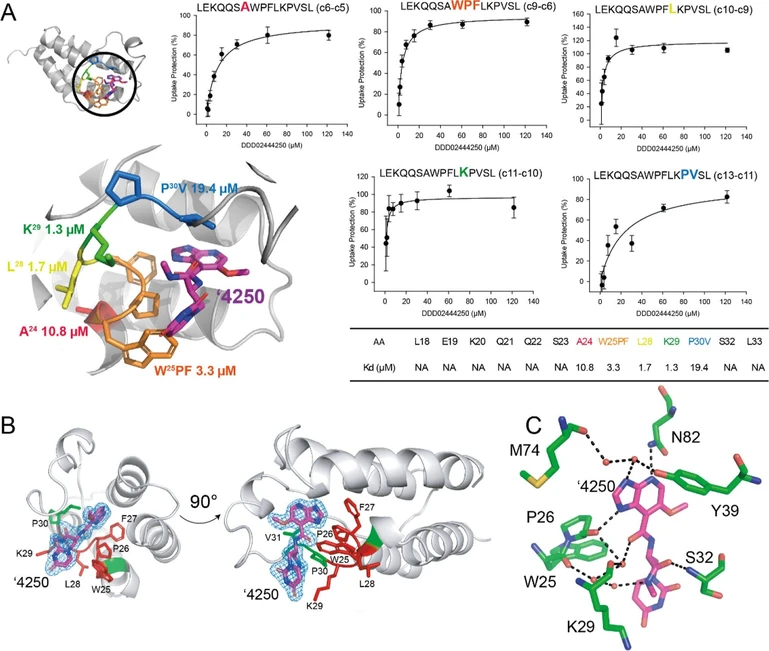 J. Am. Soc. Mass Spectrom. 2026, 37, 6, 1391–1401: Figure 4. Single amino-acid HDX-MS to derive spatial KDapp at a single residue resolution compared to crystallography data. (A) HDX-MS/MS was conducted using targeted ECD on a single peptide while titrating the ′4250 compound. Consecutive fragments are used to determine individual exchange rates, from which % deuteration can be derived. Each data point is averaged with standard deviation, from n = 3, with each exchange reaction being conducted independently. (B) Detail of ′4250 electron density binding to PfGCN5-BRD with amino acids pertinent to subsequent HDX-MS/MS analysis colored red/green. (C) Map of potential polar contacts between ′4250 and PfGCN5-BRD as identified via crystallography. Water molecules shown as red spheres.
Comparing the crystal structure of ′4250 with PfGCN5-BRD to the HDX-MS/MS there is a high degree of agreement (Figure 4B,C). There are several contacts between ′4250 and the amino acids identified in the HDX-MS/MS, although residues M74, N82, and Y39, which interact with the purine derivative part of ′4250 are not present in the HDX-MS/MS data set, we can observe that W25/K29 and S32 are present (as is the “invisible” P26). The site identified as the “tightest” interaction, K29, which had a KDapp of 1.3 μM, has a backbone hydrogen bond to a coordinated water molecule. Interestingly, there is a network of three water molecules which mediate the interaction between P26, W25, K29, and ′4250. It is possible that the presence of ′4250 slows the solvent exchange rate by stabilizing these water molecules at the site of interaction, thus slowing the solvent exchange rate. S32 was not observed to have an interaction with ′4250 via HDX-MS/MS, but the crystal structure suggests that S32’s amide group has a hydrogen bond with ′4250. The fact that this was not observed via HDX-MS/MS is interesting, as it would be expected that, given HDX measures the rate of solvent exchange on the peptide bond amide, one would expect that this residue would experience a high degree of solvent exchange rate protection on compound binding. In the “apo” structure of PfGCN5-BRD (PDB:4QNS), (43) there is no hydrogen-bonded water associated with this amide group, perhaps meaning there is no exchange event here to disrupt, or potentially, the rate of solvent exchange is slower than our 5 min time point, meaning that this interaction was outside the kinetic window of the HDX-MS/MS experiment.
Conclusion
Recent advances in peptide fragmentation technologies for HDX-MS/MS (2,17,18,44) have renewed interest in achieving residue-level localization of deuterium uptake. Here, we demonstrate that HDX-MS/MS using ECD can not only localize ligand binding sites but, through compound titration at selected exchange time points, also be used to derive apparent dissociation constants (KDapp). This represents a novel and generally applicable in-solution approach to quantifying ligand affinity without the need for protein tagging or immobilization.
HDX-MS has been used before to derive affinity constants, with studies such as PLIMSTEX (14) being capable of determining protein/ligand affinity and SUPREX (15) focusing on protein/DNA affinity. However, both of these approaches essentially measured the entire protein’s change in deuteration uptake on ligand binding, meaning that while a high-quality, in-solution affinity constant using HDX-MS could be obtained, the spatial dimension afforded by middle-down HDX-MS had been lost. Using a HDX-MS (as opposed to HDX-MS/MS) workflow, we were able to assess the local KDapp for ligand binding at a peptide level resolution. This workflow is simple, can be conducted using commercial HDX-MS systems, and offers a rapid and automatable means of determining in solution KDapp.
An interesting aspect of applying a titration-based HDX-MS approach is that it produces a distribution of apparent affinity constants across the protein sequence rather than a single value. This may seem paradoxical as ligand binding is a classical 1:1 equilibrium between a single bound and a single unbound state; (45) thus all residues in the protein should in principle report the same KD. The apparent heterogeneity in KDapp observed here, therefore, requires interpretation.
Under EX2 conditions, the deuterium uptake at any given amide hydrogen reflects a time-averaged occupancy-weighted average of the local conformational flexibility in the bound and unbound states. For residues directly contacting the ligand backbone─where binding directly suppresses amide hydrogen exchange through steric occlusion or hydrogen bonding─the ΔD signal will closely track ligand occupancy, and the fitted KDapp will most faithfully reflect the thermodynamic binding constant. For residues that are protected indirectly, through ligand-induced conformational stabilization propagated across the binding interface, the value of ΔDmax at saturation will be smaller (because the per-residue contribution of stabilization is lower), and the associated KDapp may deviate from the global value as a function of how cooperatively that stabilization is coupled to ligand occupancy. Furthermore, residues that are exchanging on time scales faster or slower than our fixed exchange time point may be systematically under- or over-represented in their apparent sensitivity to the binding event. The fact that the mean KDapp across all peptides approximates the SPR-derived KD is encouraging, but this agreement rests on a single compound–protein pair and should be tested systematically. Finally, a degree of cooperativity is expected to operate within these measurements: once the ligand has occupied the binding pocket, neighboring residues that have little direct contact with the compound are nonetheless more likely to be conformationally restrained, and this indirect effect will be reflected in their KDapp values. Further experimentation across a broader array of protein targets and binding modalities, including allosteric modalities in nonequilibrium environments (46) is required to develop a predictive framework for interpreting the spatial KD distribution in terms of the underlying interaction chemistry.
A key advantage of HDX-MS/MS is its ability to resolve affinity information at single-amino-acid spatial resolution, providing insight into local modes of interaction that complement traditional Structure––Activity Relationship (SAR) studies. The combination of HDX-MS-derived spatial interaction mapping with conventional SAR analysis can accelerate hit validation and early lead optimization by linking the compound structure to localized protein response. The lead optimization stage of drug discovery often requires a medium-throughput structural biology approach to determine how adjustments in lead compounds alter the target engagement. HDX-MS/MS is well suited to facilitate a semiautomated in-solution approach to screen these relatively small (10–30) compound series where both KDapp measurements on a peptide and amino acid level can inform likely compound engagement profiles, especially if coupled to HDX-MS/MS restraint-guided drug docking simulations.
The peptide-level KDapp determination approach described in this study is compatible with standard HDX-MS instrumentation and can be readily adapted to other protein–ligand systems, particularly where in-solution binding measurements are advantageous. The affinity measurement can also be conducted without the use of protein affinity tags (such as His/Strep-tags) or the need for fluorescent moieties, which may introduce artifacts into the experiment. Future developments in automated ECD data acquisition and analysis are expected to further improve throughput and robustness, expanding the applicability of this approach in drug discovery.


_s.webp)


_s.webp)