News from LabRulezLCMS Library - Week 15, 2025
LabRulez: News from LabRulezLCMS Library - Week 15, 2025
Our Library never stops expanding. What are the most recent contributions to LabRulezLCMS Library in the week of 7th April 2025? Check out new documents from the field of liquid phase, especially HPLC and LC/MS techniques!
👉 SEARCH THE LARGEST REPOSITORY OF DOCUMENTS ABOUT LCMS AND RELATED TECHNIQUES
👉 Need info about different analytical techniques? Peek into LabRulezGCMS or LabRulezICPMS libraries.
This week we bring you application notes by Agilent Technologies, Shimadzu, Thermo Fisher Scientific and Waters Corporation!
1. Agilent Technologies: Choosing Appropriate Pore Size Columns for Size Exclusion Chromatography of Oligonucleotides
- Application note
- Full PDF for download
SEC has become the method of choice for aggregate analysis of biotherapeutic molecules. Unlike proteins, which generally have a compact structure, oligonucleotides are much larger. They may be single- or double-stranded, and they may have a complex structure containing loops and hairpins. These structures may also vary depending on the conditions used for analysis.
This study used SEC of oligonucleotide standards available in a wide range of sizes. These sizes corresponded to differing dimensions in solution that covered the entire resolving range of the columns used in the study. This allowed a direct comparison of the chromatographic properties of each column, such as exclusion limit (rather than pore size) based on the number of nucleotides.
Although oligonucleotides are highly hydrophilic, they can exhibit secondary interactions through hydrogen bonding or from ionic interactions. Also, variations that introduce hydrophobic modifications such as labels can also cause secondary interactions. It is therefore advisable to screen different mobile phase conditions to ensure robust methodology.
Experimental
Instrumentation
Data acquisition was performed on an Agilent 1260 Infinity II bio-inert LC system using Agilent OpenLab CDS software.
Results and discussion
The first experiment was to determine if using a slower flow rate would improve chromatographic performance as very large molecules diffuse more slowly. Figure 1 shows the separation of a 50 bp DNA ladder performed at 0.175 and 0.35 mL/min. The latter is the flow rate normally used with 4.6 mm id SEC columns.
The main difference was the peak height – at the lower flow rate, the peaks spent longer in the detector flow cell, resulting in a greater response. Other factors such as peak width and resolution appeared to be very similar.
However, when testing larger oligonucleotides using the 1 kb DNA ladder (Figure 2), the peak response was similar, confirming that larger molecules have slower diffusion times. Consequently, the resolution of the largest molecules was improved, and subsequent experiments were performed using the lower flow rate. Figure 3 shows the separation of the 1 kb DNA ladder using the Agilent AdvanceBio SEC 1000 Å, 2.7 µm column with three different mobile phase solutions as described in Table 1. The total buffer/salt concentration was either 150 or 300 mM, ensuring that nonspecific secondary interactions were minimized. The molecules eluted at almost the same retention time (RT) with very similar peak shape and resolution. The 2X PBS mobile phase conditions were used for creating the calibration data plots shown in Figure 4. Individual retention times are listed in Table 4 and were determined by integration of chromatograms shown in Figure 5.
Conclusion
The columns used in this study are the latest Agilent AdvanceBio SEC columns in 500 and 1000 Å pore sizes, which provide exceptional pore volume for maximizing the useful range of oligonucleotides that can be separated. It is difficult to compare the resolving range of globular proteins to the resolving range of oligonucleotides, but a useful indication comes from the pore size of the column. For example, 500 Å pore size columns are suitable for DNA up to approximately 500 to 1,000 bp while 1000 Å pore size columns are suitable for DNA up to approximately 2,000 to 3,000 bp (Figure 8).
2. Shimadzu: Simultaneous Analysis of Synthetic Colorants, Including Red No. 3, Using an Organic Silica Hybrid Core-Shell Column
- Application note
- Full PDF for download
User Benefits
- The organic silica hybrid-based Shim-pack NovaCore C18-HB provides a wide pH tolerance range from 1 to 12 for both the mobile phase and sample solvent.
- A reduction in analysis time compared to fully porous columns can be achieved because separation on the Shim-pack NovaCore C18-HB column occurs only in the porous outer layer of the particle (core-shell column).
- LabSolutions MD, dedicated software for supporting method development, enables efficient exploration of separation conditions
Food colorants can be categorized into natural and synthetic types. The availability of synthetic colorants varies across countries, depending on regulatory frameworks. In Japan, twelve synthetic colorants are currently approved as food additives, and their safety can be assessed through qualitative and quantitative analysis using liquid chromatography. Recently, the U.S. Food and Drug Administration announced a prohibition on the use of Red No. 3, one of these twelve colorants, in food products, further increasing interests regarding the safety of synthetic colorants. This study presents the optimization of separation conditions for the simultaneous analysis of these twelve synthetic colorants using the organic silica hybrid core-shell column, Shim-pack NovaCore C18-HB, in combination with LabSolutions MD, a dedicated software for supporting method development. Furthermore, the applicability of the optimized method is demonstrated through the analysis of a real sample, Konpeito sugar candy.
About Shim-pack NovaCore C18-HB
Shim-pack NovaCore C18-HB is a core-shell column incorporating an organic silica hybrid material. Conventional silica gel is known for its low chemical stability under basic conditions, with a pH tolerance range of 2 to 7.5. In contrast, organic silica hybrid materials exhibit a broader pH tolerance, ranging from 1 to 12. As a result, the Shim-pack NovaCore C18-HB is applicable not only under acidic conditions but also in basic pH environments, making it suitable for method development processes. In coreshell particles, the inner core is non-porous, while the outer layer is porous. In contrast, fully porous particles allow analytes to diffuse deeply into the inner structure, where separation and diffusion occur. In core-shell particles, however, molecular diffusion is restricted to the thin outer porous layer, leading to more efficient separation due to reduced band broadening. This structural difference enables core-shell columns to achieve sharper peak shapes and shorter analysis times.
Conclusion
A case study was presented on the optimization of separation conditions for the simultaneous analysis of twelve synthetic tar colorants using Shim-pack NovaCore C18-HB column and LabSolutions MD, followed by the analysis of a real sample (Konpeito sugar candy). The core-shell column is constructed with an organic silica hybrid material, allowing the use of a wide pH range (from 1 to 12) for both the mobile phase and sample solvents. Additionally, it offers the advantage of reduced analysis times compared to fully porous columns.
3. Thermo Fisher Scientific: Comprehensive screening of per- and polyfluoroalkyl substances (PFAS) in food contact materials
Utilizing combustion ion chromatography fo total organic fluorine (TOF) analysis
- Application note
- Full PDF for download
PFAS testing has been performed primarily using liquid chromatography coupled with triple quadrupole mass spectrometry (LC-QQQ). As a targeted analytical technique, LC-QQQ results are limited to compounds with available standards. As a result, these targeted studies do not necessarily provide a comprehensive measurement of the total PFAS that may exist in samples. Recently, laboratories have focused on developing and validating lower-cost alternatives that provide a more comprehensive measure of total PFAS content. This has led to the development of several methods for measuring total fluorine (TF) as a proxy for total PFAS contamination in FCM. These methods employ technologies such as combustion ion chromatography (CIC)10-13 and particle-induced γ-ray emission spectroscopy (PIGE).14-16 PIGE is a surface measurement technique. Due to the limited penetration depth of the particle beam, sample heterogeneity and surface coatings can lead to higher measurements compared to bulk volume techniques such as CIC. However, measuring only TF is not a reliable proxy for PFAS, as it includes both organic and inorganic fluorine, the latter of which is not considered PFAS. Using TF as a measure of PFAS may overestimate the amount of PFAS in samples. A method for determining TOF in packaging was developed using oxygen combustion sample preparation with a fluoride ion-selective electrode.17 However, this method involves tedious manual steps and lacks the sensitivity of CIC. Alternatively, some studies have used extractable organic fluorine (EOF) to better indicate the total amount of PFAS in samples.10-12 However, these studies provided a screening technique for PFAS with CIC without specifying the type of fluorine extracted (inorganic vs. organic). This can overestimate EOF if inorganic fluorine (IF) is not subtracted from the measurement, as there is the potential for co-extraction of IF using methanol and other polar organic solvent.18 Additionally, previous studies only cut FCM into small pieces without grinding them into powder, likely resulting in lower extraction efficiency compared to samples ground to increase the surface area for extraction.
CIC offers excellent sensitivity and versatility, independence on sample thickness, and the possibility for direct ion chromatography (IC) analysis to determine IF. Consequently, CIC has been successfully used to determine adsorbable organic fluorine (AOF) and TOF in environmental sample matrices.19-21
In this study, we developed a method to screen for total PFAS using CIC to characterize fluorine content in FCM. This method allows for full quantification of TOF and EOF fractions of fluorine. Furthermore, we characterize the organic fluorine fractions by identifying individual PFAS compounds using high resolution mass spectrometry (HRMS).
Experimental
Equipment
A Thermo Scientific™ Dionex™ Integrion™ HPIC™ system (P/N 22153-60306) including:
- Eluent generator
- Pump
- Degasser
- Conductivity detector
- Column oven temperature control
- Detector-suppressor compartment temperature control
Nittoseiko™ Automatic Combustion Unit Model AQF–2100H system* including:
- Automatic Sample Changer ASC-Controlasc-270LS
- Horizontal Furnace Model HF-210
- Gas Absorption Unit GA-211
- External Solution Selector ES-210
*Any combustion oven with equivalent performance will work
Thermo Scientific™ EXTREVA™ ASE™ Accelerated Solvent Extractor (P/N 22184-60101)
Genevac™ Rocket Synergy™ 2 Benchtop Evaporator (ATC Scientific Product, Warminster, PA)
Thermo Scientific™ Orbitrap Exploris™ 240 mass spectrometer (P/N BRE725535)
Thermo Scientific™ Vanquish™ Flex UHPLC system
Software
- All CIC data were acquired using the Thermo Scientific™ Chromeleon™ Data System (CDS) Version 7.3 1 with DDK driver to control the combustion system.
- All LC-HRMS data were acquired and processed using Chromeleon CDS, version 7.3.2.
- Thermo Scientific™ Compound Discoverer™ 3.3 SP3 software package
Results and discussion
Identification of PFAS in FCM using HPLC-HRMS
The levels of total organic fluorine (TOF) detected in the three samples exceed the regulated limit for PFAS in food contact materials (>100 ppm). Sample #1 also surpasses the regulated limit of 100 ppm for extractable organic fluorine (EOF). To better understand the specific molecular composition of the EOF fraction collected from FCM food contact materials, a nontargeted analysis approach using LC-HRMS was implemented for extracts of all three food contact material samples. Following recommendations from Charbonnet et al., specific criteria were established to classify the annotations of each detected PFAS compound into five confidence levels (Table 3). Confidence Levels 1 and 2 require strong matches to mass spectra within libraries of spectra obtained from authentic reference standards. Level 3 necessitates a combination of high matching scores to spectra within an in silico mass spectra library or a PFAS fragment database. Annotations in Levels 4 and 5 rely on a match between the measured monoisotopic mass (within 2 parts per million) and/or a high similarity between the measured and hypothetical isotope pattern.
By analyzing the relative peak area intensities, it was found that a significant number of the most abundant PFAS compounds belonged to a homologous series of perfluorocarboxylic acids (PFCAs) ranging from perfluoropropionic acid (C3; PFPrA) to perfluorooctanoic acid (C8; PFOA). All of these PFCAs except one (perfluoropropionic acid) were confidently annotated at Level 1 and observed in all three samples. PFOA, however, was only detected in the paper bowl and paper plate. The cookie bag contained the highest total summed peak areas for all PFCAs, followed by the paper plate and the paper bowl. Previous studies have also reported PFCAs in various food contact materials.
In addition to the linear PFCAs, a perfluorinated cyclic isomer of perfluorocarboxylic acids, perfluoro methyl cyclopentane carboxylic acid, was detected in all three samples, with the highest peak area observed in the cookie bag. Furthermore, perfluorobutanesulfonic acid (PFBS) was detected in all three samples, with the paper bowl having the highest abundance. Both PFBS and the PFCAs were confidently annotated at Level 1. 14
In addition to perfluorinated compounds, several polyfluorinated compounds were detected and confidently annotated at Levels 3-5. This includes 2-perfluorohexyl ethanoic acid (6:2 FTCA), 3:2 fluorotelomer alcohol (3:2 FTOH), and 6:2 fluorotelomer sulfate (6:2 FTS), all of which were found in all three samples. Among them, 6:2 FTCA exhibited the highest peak area response in the cookie bag, while 3:2 FTOH and 6:2 FTS were most prevalent in the paper bowl. Previous research has reported the presence of 6:2 FTCA and 6:2 FTS in food contact materials. Fluorotelomer alcohols (FTOH) like 6:2 FTOH and 8:2 FTOH have also been documented in prior studies.2,3 However, the observation of 3:2 FTOH in this study is novel to the best of our knowledge.
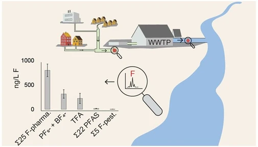
When considering the total peak areas of different species within major homologous groups (perfluorocarboxylic acids, PFCAs; perfluorosulfonic acids, PFSAs; fluorotelomer alcohols, FTOHs; fluorotelomer carboxylic acids, FTCAs), PFCAs were found to be the most abundant group, accounting for 27–41% of the total peak area of all detected compounds (Figure 7). Across all samples, these four groups collectively represented 50–63% of the total peak areas of all detected compounds.
Conclusion
We developed a sensitive and automated method using CIC to determine both TOF and EOF in FCM. The TOF method is beneficial for manufacturers to comply with current state regulations. Interestingly, we found that the amount of extractable fluorine in FCM is limited, indicating that targeted LC-MS approaches may overlook a significant portion of the fluorinated content in the samples, including unidentified EOF compounds and non-extractable organic fluorine. To address this limitation, we employed a non-targeted LC-HRMS approach, which allowed us to detect 46 PFAS compounds in the EOF fraction, many of which fall outside of the typical list of targeted screening or quantification methods. Among these compounds, perfluorocarboxylic acids emerged as the most abundant homologous group. These findings demonstrate that this CIC workflow provides a more comprehensive understanding of the total PFAS and fluorinated content in FCM compared to LC-MS targeted approaches, offering greater clarity about the PFAS contamination in FCM.
4. Waters Corporation: Parallel Column Regeneration for Increased Analytical Throughput of Serum Steroid Hormones in Clinical Research
- Application note
- Full PDF for download
Benefits
- 18% increase in throughput of steroid hormone analysis compared with single channel separations
- Efficient shared calibration approach demonstrates equivalence with single channel analysis
Extended gradient chromatographic separations are sometimes needed to resolve matrix and isobaric interferences when analyzing complex mixtures by liquid chromatography, Tandem Mass Spectrometry (LCMS/MS). This can create a challenge for clinical research laboratories striving to meet throughput demands.
Throughput can be limited when routine, single column gradient LC analyses use one pump to load and separate one sample at a time, with data acquisition running in a series that cannot progress until the column has completed a wash and re-equilibration (regeneration) cycle. This approach can be made more efficient by taking the active column offline and using a second LC pump for regeneration. A second column can then be brought online for loading, separation and analysis of the next sample, while the first column is regenerating offline. This approach is termed ‘parallel column regeneration.
Here, we demonstrate the time-savings achieved when parallel column regeneration is applied with the rapid clinical research method for the quantification of androstenedione (A4), testosterone (T), 17 hydroxyprogesterone (17OHP), dihydrotestosterone (DHT), dehydroepiandrosterone (DHEA), and progesterone (P) in human serum using LC-MS/MS. The validity of applying a single calibration curve to results generated on both columns is explored.
The test system, an ACQUITY™ UPLC™ I-Class PLUS SM-FL BSM/BSM Parallel Column Regeneration System with Single Elution Pumps (p/n: 176005409), is shown in Figure 1. Analytes were detected and quantified in the eluted samples using a Xevo™ TQ-S Micro Tandem Mass Spectrometer, operating in positive electrospray ionization mode, and with multiple reaction monitoring (MRM) acquisitions.
Results and Discussion
The correlation coefficient of the linear regression of the calibrators through method verification was ≥0.999 and ≥0.997 (3 s.f.) for single, and parallel column regeneration, respectively (n=5 analyses). Refer to certificates of analysis for p/n: 186009311IVD for details of ranges covered by the MassTrak Endocrine Calibrator set.
Separating the detection and regeneration phases across two channels reduced the injection cycle time from 7.2 to 6.1 minutes per sample, which equates to an analysis time saving of 1.8 hours for a full 96-well plate of prepared samples.
Taking regeneration offline also allowed increased column flushing and equilibration (Figure 3), potentially removing more residual sample matrix from the LC column, and giving the opportunity to create optimal conditions for robust and stable chromatographic performance of early eluting, relatively less-retained analytes.
Single and parallel column imprecision was acceptable at ≤15% relative standard deviation (RSD), with the exception of single column analysis of dihydrotestosterone (DHT) in low concentration control samples. Interestingly, the within-batch and total precision was improved for this analyte at low concentrations, when using parallel column regeneration. A larger sample size is needed, however, to draw firm conclusions regarding the statistical significance of any effects on variance.
The results of EQA sample analysis suggested no significant quantitative differences between single and parallel column regeneration, and this was confirmed in the longer term, with the finding that mean control sample results made with parallel column regeneration were not significantly different to single column results (Student’s t-test, data not shown, p<0.05). Samples for DHEA EQA were not available for analysis. Splitting calibrators between two LC columns gave similar results to those derived from a full set of calibrators analyzedanalysed using a single column. The potential to apply a single calibration across two LC columns presents an efficient and simplified acquisition and processing workflow.
Conclusion
Parallel column regeneration increased the efficiency of sample analysis without compromising column care and use best-practices. This technique may be of interest to any laboratory involved in the analysis of large numbers of clinical research specimens.