Tracking Protein Misfolding and Oligomerization: A Temperature-Controlled Ion Mobility-Mass Spectrometry Approach
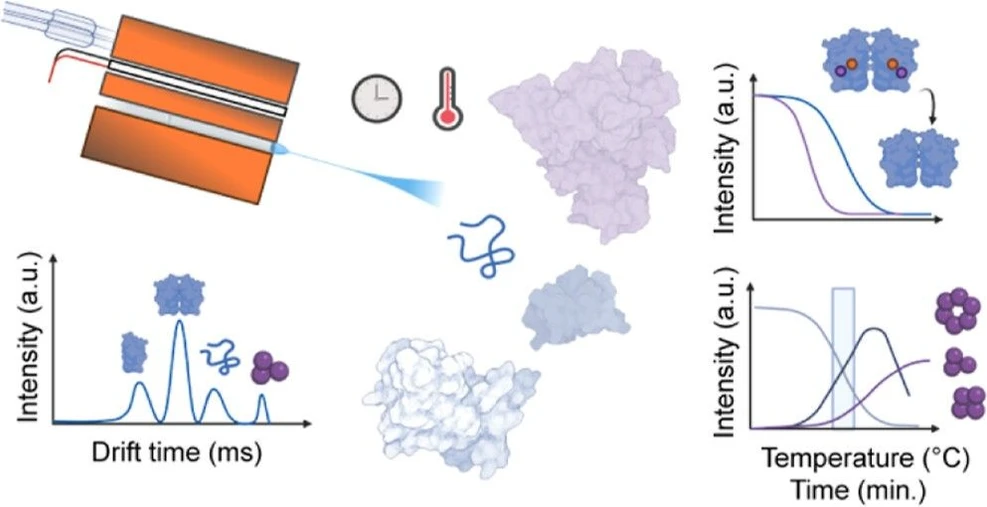
Anal. Chem. 2026, 98, 20, 14683–14694: Graphical abstract
This study introduces a temperature-controlled nanoelectrospray ionization ion mobility–mass spectrometry (TC-nESI-IM-MS) workflow for investigating protein misfolding and oligomerization. Combined with surface-induced dissociation and limited proteolysis, the approach enables real-time characterization of transient folding intermediates and early aggregation events in Cu/Zn superoxide dismutase (SOD1).
The results revealed distinct unfolding and oligomerization pathways for holo- and apo-SOD1, identifying key structural regions involved in non-native oligomer formation. The platform provides detailed mechanistic insight into early protein aggregation and offers a powerful analytical strategy for studying molecular processes underlying neurodegenerative diseases.
The original article
Tracking Protein Misfolding and Oligomerization: A Temperature-Controlled Ion Mobility-Mass Spectrometry Approach
Despoina Svingou, Luke McAlary, Julian Alexander Harrison*, and Renato Zenobi*
Anal. Chem. 2026, 98, 20, 14683–14694
https://doi.org/10.1021/acs.analchem.5c06100
licensed under CC-BY 4.0
Selected sections from the article follow. Formats and hyperlinks were adapted from the original.
Neurodegenerative diseases encompass a variety of disorders characterized by the progressive disruption of the structure and function of neurons. (1−3) These debilitating diseases, exhibit a complex mechanism of aberrant protein misfolding, oligomerization, aggregation and ultimately amyloid plaque formation. (2,4) A major hurdle in elucidating misfolding and early aggregation of proteins has been the lack of techniques to capture and analyze low-abundance intermediates taking part in this dynamic process in real time. (5) Traditional methods, which mostly yield global information and require long incubation times, often fall short in providing the temporal and structural resolution necessary to study these dynamic and conformationally diverse systems. (5,6)
Oligomeric complexes are commonly studied by spectroscopic methods such as single-molecule fluorescence and nuclear magnetic resonance (NMR) spectroscopy, by size-exclusion chromatography (SEC), or by atomic force microscopy (AFM). (7,8) However, these low-abundance and transient oligomeric assemblies, often coexist in solution, which presents a significant challenge for traditional biophysical techniques. While powerful, these methodologies typically provide global information rather than unambiguous structural and conformational characterization that would be needed to resolve dynamic oligomeric ensembles. (9)
One technique that has the potential to overcome these challenges is native mass spectrometry (MS). This powerful tool can be used for the characterization of protein complexes and the biophysical properties of their interactions. (10−12) In native MS, soft nanoelectrospray ionization (nESI) and optimized gas pressures and voltage conditions maintain delicate molecular interactions of even large noncovalent complexes in the gas phase, allowing for detailed structural analysis of heterogeneous protein complex ensembles. (13,14) Even though structural rearrangements are expected upon desolvation, proteins have been shown to retain many structural aspects upon transfer to the gas phase. (15) Thus, native MS can serve as a valuable technique that is directly correlated to solution-based methodologies.
Additionally, important information about the conformations adopted by copopulated biomolecular complexes can be derived when native MS is coupled to ion mobility spectrometry (IM-MS), where the overall shape of the ions and their rotationally averaged collision cross section (CCS) can be determined based on their mass, charge and interactions with the drift gas. (16,17) Especially the development of high-resolution IMS platforms shows great promise in deciphering the conformational landscape of proteins. (18,19) Another methodology, known as variable-temperature or temperature-controlled nESI (TC-nESI), offers an avenue to probe such processes by accelerating aggregation under defined thermal conditions before ionization. (20,21) When integrated with high resolution IM-MS, it can provide valuable insights into the thermodynamic properties, stability, folding and interactions of protein complexes. (22−24) In this study, we explore its potential to induce and access previously unexplored aggregation pathways in real time.
The multitude of pathways a neurodegenerative disease-related protein can take is exemplified here by Cu/Zn superoxide dismutase (SOD1), as it encompasses several molecular characteristics of amyloidogenic proteins, including conformational plasticity, misfolding and metal-regulated stability, folding and assembly (Figure 1). (25−28) SOD1 is a 32 kDa homodimeric protein that catalyzes the conversion of superoxide, to either H2O2 or O2, depending on the Cu-oxidation state. Each of its monomers is characterized by a β-barrel scaffold consisting of eight antiparallel β strands and two loops (an electrostatic and a zinc-binding loop). The dimer also contains one copper and one zinc ion per 153-residue subunit. (29,30) In its fully metalated state, the SOD1 dimer is referred to as holo-SOD1, whereas the demetalated species is known as apo-SOD1. Currently, over 200 SOD1 gene mutations are implicated in amyotrophic lateral sclerosis (ALS). (31,32) These mutations can affect the structure and stability of the protein, causing dissociation of metal cofactors, the formation of transient neurotoxic oligomers, and finally large insoluble inclusions, all hallmarks of neurodegenerative disease. (33,34)
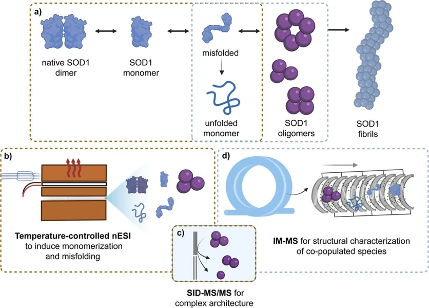 Anal. Chem. 2026, 98, 20, 14683–14694: Figure 1. Schematic of methodologies employed to characterize the stages of SOD1 aggregation from native dimer to insoluble fibrils. (a) The SOD1 aggregation pathway proceeds from native dimers to conformationally diverse monomers and subsequently to soluble heterogeneous oligomers, prior to insoluble fibril formation. (b) SOD1 dimer dissociation and monomer misfolding can be induced by controlled thermal ramping using TC-nESI (brown boxes). (c) The architecture of oligomeric complexes can be determined by SID-MS/MS. (d) IM-MS featuring the cyclic IMS design enables structural and conformational characterization of all species (blue boxes).
This work aims to establish a MS-based workflow for probing the early stages of misfolding and oligomerization in neurodegenerative disease–related proteins (Figure 1a). The approach enables precise control of aggregation speed and pathways through temperature-controlled nanoelectrospray ionization (TC-nESI) (Figure 1b). Resulting oligomers are characterized using high-resolution IM-MS, surface-induced dissociation (SID), and limited proteolysis, providing detailed structural and conformational insights at each stage (Figure 1c–d). This generalizable methodological framework allows the investigation of previously uncharacterized intermediates, offering critical understanding of the molecular mechanisms underlying disease progression and aiding in the development of therapeutic strategies.
Materials and Methods
Native IM-MS Experiments
All native IM-MS experiments were carried out on a SELECT SERIES Cyclic IMS (Waters, Wilmslow, U.K.), equipped with a quadrupole filter that allows isolation of species up to 32000 m/z, followed by a three-lens SID cell placed in front of the trap collision cell. (36,37) All samples were sprayed in positive mode from borosilicate glass capillaries (inner diameter of ∼1 μm) that were pulled in-house employing a micropipette puller (P-1000, Sutter Instruments) and fitted with a platinum wire. The mass range was set from 50 to 32000 m/z for the initial holo-SOD1 study to accommodate higher-mass oligomers and was modified to 50–16000 m/z for the rest of the experiments. For the heating experiments with a ramp of 1.0 °C min–1, the scan rate (i.e., the time over which each spectrum was collected) was set to 1s. When a heating rate of 0.3 °C min–1 was employed the scan rate was changed to 2s, to reduce data size during longer acquisitions. For all experiments the mass spectrometer was operating in “V-mode”, while all data were acquired in “Mobility mode”. Primarily, crucial cIMS parameters for high-mass ion transmission and optimal ion mobility separation were considered, as described by Harrison et al. (11) Operating parameters for all experiments: native IM-MS and collision-induced unfolding tandem MS (CIU-MS/MS), are described in the Supporting Information (Tables S1–S4).
Data Acquisition and Processing
All IM-MS data were acquired using MassLynx 4.2 (Waters). Extraction of retention time and drift time profiles, as well as smoothing of acquired spectra, were conducted using Driftscope v3.0 software (Waters) and MassLynx 4.2 (Waters), respectively. Deconvolution of SID-MS/MS data was achieved using UniDec software. (42) Further data analysis and visualization were performed using Prism 10.1.1 (GraphPad Software) and Adobe Illustrator (26.0.3, Adobe Inc., California). Graphics were created using Biorender.
All thermal denaturation results represent summed data across the entire temperature ramp, unless stated otherwise. Additionally, all melting and formation temperatures reported are defined as the midpoints of sigmoidal or reverse sigmoidal abundance curves for each species, respectively.
Results and Discussion
Method Development and Optimization
To showcase our approach, bovine SOD1 was chosen as a model system due to its high similarity to human SOD1 (82% sequence similarity) and its well-characterized folding behavior, making it an ideal platform to benchmark early aggregation studies. (48,49) Here, thermal denaturation MS experiments along with high-resolution ion mobility separation, were employed for the simultaneous characterization of thermal stability and oligomer growth mechanisms. However, several methodological adaptations need to be made to study this system with temperature-controlled mass spectrometry. Thus far, most studies using this technique employ heating rates ranging from 1.0 or 2.0 °C min–1 to much faster thermal ramping of laser-based methods, to probe solution-phase stability and thermodynamics. (11,22,50,51) However, the typically used heating rates may not be suitable for studying proteins involved in neurodegenerative disease due to the intricate kinetics of intermediate formation in such systems. (52) Therefore, we explored different heating rates for a range of temperatures, focusing on their effect on the interplay of SOD1 native dimer dissociation, unfolding and oligomer formation.
Initially, holo-SOD1 was subjected to heating at a rate of 1.0 °C min–1 in the temperature range of 25–87 °C (Figure 2). At the beginning of the ramp, the main ions in the spectrum corresponded to the native dimer, with charge states ranging from 9+ to 12+, with 11+ being the most prominent (Figure S2). Interestingly, for the main charge state of the native dimer (211+), three distinct conformations were observed, with CCS of 2826 (native), 3334 and 3666 Å2 consistent with partial unfolding of the dimeric structure (Figure S3). The latter two conformers are of low abundance and cannot be identified in the 10+ and 12+ charge states due to overlap with the 5+ and 6+ monomers, respectively (Figure 2). (26) Other species observed, in lower abundances, include monomer (in charge states ranging from 5+ to 8+) and tetramer (ranging from 14+ to 17+). Only one conformational population was initially observed for both (Figure S2).
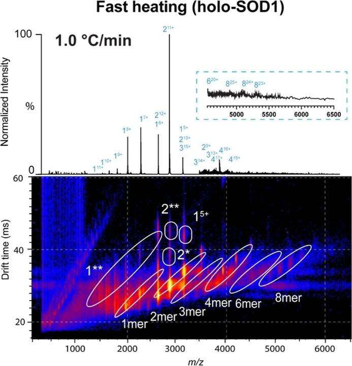 Anal. Chem. 2026, 98, 20, 14683–14694: Figure 2. Fast thermal denaturation of holo-SOD1 via TC-nESI-IM-MS. Mass spectrum of holo-SOD1 after fast heating (data summed over the range of 75–85 °C). The data were acquired with a rate of 1.0 °C min–1 over the full 25–85 °C ramp. The zoomed-in spectrum shows the m/z region from 4500 to 6500. The lower m/z range (50–2300) is x10 magnified, while the higher m/z range (4000–6500) is ×200 enhanced. The corresponding heat map shows three-dimensional data of the fast ramp offering intensity, drift time and m/z of all species. The spectra from both the fast and the slow ramp (Figure 3a) contain the same number of scans.
For the fast-heating experiment, at 75 °C, holo-SOD1 first dissociates from dimers into monomers (Figure 2). Throughout this work, misfolded intermediates are denoted as (*), and fully unfolded species as (**), as determined by their charge state and experimentally derived CCS. The IM-MS profile indicated that the monomers formed two populations (Figure 2). Based on CCS and charge state, these species corresponded to folded (1) and unfolded (1**) monomers (Figure S4). The simultaneous appearance of two monomeric populations could stem from localized monomer unfolding prior to dimer dissociation, a phenomenon unlikely in solution and previously attributed to gas-phase dynamics. (53) However, as the abundance of those populations increased by heating, they can be considered dimer dissociation products resulting from increased internal energy within the complex (Figures 2 and S5). This can yield monomers of variable degrees of unfolding, with the prospect of partial or complete metal loss. Concerning higher-order oligomeric structures, mainly tetramers were observed, along with some very low-abundance trimers, hexamers and octamers (Figure 2). The preliminary data for the typical ramp prompted two prescient questions: can the formation of oligomers be better captured using different heating parameters? How is the monomer folding population related to oligomer assembly? To explore how the kinetics of monomer formation and misfolding influence SOD1 oligomer assembly, we modified the heating rate of the temperature ramp for this aggregation assay.
Investigating the Assembly Mechanism of Apo-Oligomers
Investigating the architecture of transient and low-abundance heterogeneous oligomeric species at the brink of large aggregate formation is inherently challenging. The difficulty increases especially when trying to probe large dynamic structures in the gas-phase, while maintaining solution-phase structural fidelity. To address this, we employed a multifaceted approach combining surface-induced dissociation tandem mass spectrometry (SID-MS/MS) and limited proteolysis at elevated temperatures. This strategy allows access to both solution-phase and gas-phase interactions through proteolytic cleavage, heat-induced fragmentation, and stepwise complex dissociation, respectively. (65) In doing so, we gain valuable insights into oligomer formation mechanisms and identify potential labile regions that may serve as oligomerization interfaces.
First, SID-MS/MS experiments were performed after a slow temperature ramp (55–60 °C, 0.3 °C min–1), ensuring oligomer formation. A wide range of surface collision energies from 20 to 110 V was explored for all oligomeric assemblies, and relative intensities of precursor and dissociation products along this range of potential were traced. The results of the dissociation pathways of each oligomer are depicted in Figure 5, where the cartoons illustrate the most probable dissociation pathway. In such an investigation, quadrupole selection of specific charge states is challenging, as there is significant overlap from different oligomers within the same m/z window. As charge state selection can dictate charge partitioning and fragmentation efficiency in SID, lower charge states are expected to provide more information about the architecture, retaining the structures of bigger subcomplexes. (61,66) Hence, for each complex, the lowest of the nonoverlapping charge states was chosen and subjected to SID. The exception was the tetramers, where the 17+ charge state was chosen because lower charge states showed peak overlap with other subunit configurations in the spectrum.
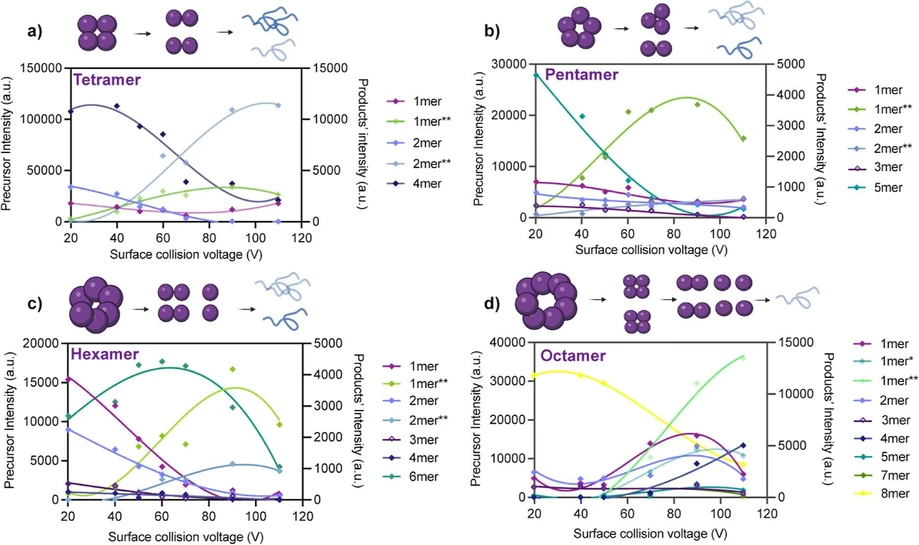 Anal. Chem. 2026, 98, 20, 14683–14694: Figure 5. SID profiles of different apo-oligomers. (a–d) Apo-oligomers investigated range from tetramers to octamers. The intensities of all ejected complexes (extracted from their respective IM-MS heat maps) throughout the SID-MS/MS experiment were plotted over collision surface voltage (20 to 110V). Raw intensities of precursor and product ions are traced on the left and right y-axis, respectively. The profiles of each population are color-coded to match Figure 4f. Schematics illustrating the most probable dissociation pathway each apo-oligomer are shown above the corresponding plots. In these schemes, the color intensity of each subcomplex reflects its relative abundance, while noncircular shapes represent unfolded species.
Conclusions
Ion mobility-mass spectrometry is a powerful technology that can unambiguously characterize conformationally diverse copopulated oligomeric protein complexes. This offers unique insights into the events preceding and occurring during early protein aggregation that are thought to be the most important regarding neurodegeneration. However, inducing aggregation often involves long incubation times and a combination of methods to structurally and temporally characterize an ensemble of low-abundance and transient early aggregate formations. In this work, TC-nESI was combined with IM-MS to structurally and kinetically characterize intermediates and oligomeric complexes of bovine SOD1, produced upon heat-accelerated aggregation.
For holo-SOD1, dimer dissociation to monomers of variable folding states initiated oligomerization with fast incorporation of mainly dimers, tetramers and monomers into higher-order assemblies. Interestingly, slower thermal ramping produced more misfolded monomeric SOD1 and enabled more abundant oligomer formation. Apo-SOD1 yielded more abundant and more compact oligomers, and their assembly was more gradual compared to the holo-analogue. Finally, SID-MS/MS experiments revealed multiple dissociation pathways and tetrameric, trimeric, dimeric and monomeric building blocks comprising the scaffold of apo-oligomers, while insights from limited proteolysis and heat-induced fragmentation identified loops III, V, VI and VII, and the C-terminus, as labile regions in misfolded apo-SOD1 monomers. These observations reveal a mechanism involving early native dimer dissociation, monomer misfolding and reassociation to higher-order globular oligomers via hydrophobic interactions localized at defined regions. Additional intermolecular interactions, such as disulfide bridges, between dimers are also suspected to stabilize multimers.
Overall, this study provides the first real-time, high-resolution characterization of early SOD1 misfolding and oligomerization intermediates under controlled thermal stress. By combining TC-nESI with high-resolution IM-MS, SID, and limited proteolysis, we uncover conformationally distinct monomeric and previously unobserved oligomeric species, map labile structural regions driving aggregation, and reveal how metal cofactors modulate complex assembly. The methodology developed in this work establishes an experimental framework to elucidate the structural dynamics of other amyloidogenic proteins, such as mutated human SOD1, amyloid-beta or a-synuclein. Their aggregation pathways often involve metalation-dependent assembly and misfolded monomeric intermediates, making them particularly amenable to this approach. (25,26,70,71) This strategy helps to bridge a critical gap in understanding the earliest stages of neurodegenerative protein aggregation.